Thursday 25 June 2015

Endometriosis


Definition

Endometriosis can be defined as presence of endometrial tissue outside the uterus. These tissues are also called endometrial implants or ectopic endometrial tissues.

Commonly, endometriosis / ectopic endometrial tissues are found in the following places: 1)Peritoneum, 2)Ovaries, 3)Fallopian tubes, 4)Outer surface of uterus, bladder, ureters, intestine and rectum, 5)Pouch of Douglas (rectouterine pouch).


Pathogenesis

There are few theories that has been suggested as the underlying mechanism on how endometriosis can occur, including 1)theory of retrograde menstruation, 2)coelomic-metaplasia theory, 3)circulation and implantation theory.

Pathophysiology

Ectopic endometrial tissues have similar features with the endometrial tissue lining the uterus. Just like the endometrial lining of the uterus, ectopic endometrial tissues also undergo cyclical changes of the menstrual cycle and respond to changes of the estrogen level. As we know, menstruation is associated with inflammatory process. These repetitive inflammatory events eventually lead to scarring and adhesion. All these inflammation, scarring and adhesion lead to the clinical signs and symptoms of endometriosis and its complications, depending on where is the location of the ectopic endometrial tissues.

Woman with endometriosis may be presented with symptoms of abdominal/back pain, dysmenorrhea, dyspareunia, dysuria, or dyschezia. Some women may be presented with the complications such as chronic pelvic pain and subfertility.

Diagnosis

Endometriosis should be suspected when a woman in her reproductive age presented with severe dysmenorrhea, which is not improved with NSAIDs. On physical examination in women with endometriosis, there may be pelvic tenderness, or nodularity of the uterosacral ligaments and rectovaginal septum, and ultrasound may reveal ovarian cyst and endometrioma. Laparoscopic examination, which is the gold standard procedure to confirm the diagnosis, might be done and the suspected lesions are removed and sent for biopsy. However, laparoscopy for diagnosis is not necessary to be done before starting treatment with medications. Laparoscopy should generally be performed only when the surgeon plan to remove the lesions if endometriosis is discovered during the procedure.

Treatment

Treatment modalities in the management of endometriosis can be divided into medical treatment and surgical treatment. A woman with endometriosis may be treated medically or surgically or both. The aim of the treatment should be directed to improve patient’s symptoms and quality of life, and should be tailored based on the severity of the disease and patient’s fertility desire.

Monday 22 June 2015

Procedures / active intervention in twin to twin syndrome.

There are 4 procedures which have been known to have role in the management of TTTS. The procedures include: 1) serial amniotic reduction 2) microseptostomy 3) selective fetal laser photocoagulation and 4) fetal cord occlusion / coagulation.

Amnioreduction

Amnioreduction is a procedure whereby a large volume of amniotic fluid is removed from the amniotic sac by amniocentesis procedure. A needle is introduced into the amniotic sac percutaneously through the abdomen into the amniotic sac of the recipient twin who develops polyhydramnios. The aim of amnioreduction in TTTS is to control polyhydramnios in hope of prolonging the pregnancy to reduce the risk of extreme prematurity. In addition to that, amnioreduction improves utero-placental blood flow, most likely by reducing intrauterine pressure. However, the procedure doesn’t treat the cause of the disease. The effect of amnioreduction is only temporary and the polyhydramnios may develop again as the disease progress. In order to be effective, the procedure need to be done repeatedly - ‘serial amnioreduction’. Since it is an invasive procedure, it poses risk of infection towards the pregnancy. The risk of intrauterine infection is increase with the number of repeated invasive procedure done.
amnioreduction done by amniocentesis


Microseptostomy

It is done by creating a small hole on the intertwine membrane. Similar to amnioreduction, the aim is to control polyhydramnios by amniotic fluid dynamic equalization between both amniotic sac of the recipient and donor twin. Unlike amnioreduction, this procedure doesn’t need to be repeated. Therefore, the risk of intrauterine infection is lowered compared to serial amnioreduction. However,there is risk of the hole become larger. If the hole get larger, instead of only the amniotic fluid able to cross the septostomy, the other structure, for example the cord, may as well cross the septostomy and this increase the risk of cord entanglement. 
microseptostomy


Fetal laser coagulation

This is the 1st procedure aims to treat the cause of the disease. Fetal laser coagulation therapy is a procedure whereby blood vessels on the intertwine membrane are occluded by laser therapy, thus arrests the shunting of blood from donor to recipient and stop the transfer of potential vasoactive mediators. Initially, the procedure include coagulation of all vessels on the intertwine membrane, including the normal vessels. This may lead to acute placental insufficiency since the normal vessels that carry nutrient and blood supply are also coagulated. After that, the procedure has been improved to selective fetal laser coagulation, whereby only the vessels that lead to unbalanced transfusion are occluded. This procedure considered as the most superior therapy for TTTS, however, it only available in selected institution and requires intensive training. Besides, in cases of the affected vessels lies deep in the membrane, it might not be detected thus fetal laser coagulation could not be done. 
fetal laser coagulation


Fetal cord occlusion

This procedure sacrifices one twin to save the other twin. It is the final resort for cases which there is already demise or imminent demise of one of the twin. It doesn’t only stop the progression of the syndrome, but also prolonged the gestation and maximize the outcome of the other twin.
fetal cord occlusion


Monday 15 June 2015

Quintero staging system - twin to twin transfusion syndrome


Stage
Parameter
Criteria
1
MVP of amniotic fluid
MVP<2cm in donor and MVP>8cm in recipient

2
Fetal bladder
Fetal bladder in donor twin is not visualized over 60min observation

3
Doppler waveforms:                         
At least one of these 3 abnormalities:

a-umbilical artery
Absent or reversed diastolic flow.
b-ductus venosus
Reversed a-wave flow.
c-umbilical vein
Pulsatile umbilical vein flow.

4
Fetal hydrops
Hydrops in one or both twins

5
Fetal cardiac activity
Absent cardiac activity – fetal demise in one or both twins.


Quintero system classified the disease into 5 stages based on the ultrasound findings. 5 parameters are assessed to stage the disease. The parameters include: 1) maximum vertical pocket (MVP) of amniotic fluid, 2) fetal bladder, 3)Dopplers abnormalities of the umbilical artery or ductus venosus, or umbilical vein, 4)fetal hydrops and 5)fetal cardiac activity.

Stage 1 disease occurs when the is only discordance in the amniotic fluid volume, the 1st criteria, which is detected by comparing the measurement of the maximum vertical pocket of the amniotic fluid in each amniotic sac of the donor twin and the recipient twin, with the absence of the other 4 criteria. When maximum vertical pocket of the amniotic fluid in donor sac less than 2cm and maximum vertical pocket of the amniotic fluid in recipient sac is more than 8cm, the disease is at least at stage 1. 

The presence of 2nd criteria that is non-visualization of donor twin bladder indicates at least stage 2. The disease is at least stage 3 when there is presence of the 3rd criteria that is the presence of any of these dopplers abnormalities: 1) absent or reversed diastolic flow of the umbilical artery, 2) reversed of a-wave flow of the ductus venosus or 3)pulsatile flow of the umbilical vein. Fetal hydrops (4th criteria) indicates at least stage 4 and when there is fetal demise (5th criteria) the disease already at stage 5.

-Maximum vertical pocket measurement-


-umbilical artery, ductus venosus and umbilical vein shown-

hydrops - ascites, pleural effusion and scalp edema shown-

Sunday 14 June 2015

Twin to twin transfusion syndrome - diagnosis and mangement overview

Twin to twin transfusion syndrome is diagnosed with antenatal ultrasound scan. There are 2 criteria required to diagnose the condition: 1) the presence of monochorionic diamniotic pregnancy, and 2) the presence of amniotic fluid volume discordance, i.e polyhydramnios of the recipient, defined as MVP>8cm, while oligohydramnios of the donor, with MVP<2cm.

Once a pregnancy is diagnosed with the condition, the next step is to stage the disease. The most commonly used staging system for this condition is Quintero staging system, which is based on the sonographic findings. Although it may not determine the prognosis for perinatal survival, it appears to be a useful tool to diagnose the condition, describe severity, guide treatment and monitor progression of the condition and effectiveness of the treatment.

Quintero system divides the condition into 5 stages, ranging from the most mild disease (with only amniotic fluid volume discordance) to the most severe one (with demise of one or both fetuses). Amniotic fluid volume discordance, with MVP<2cm in donor sac and MVP>8cm in recipient sac, defines stage 1 of the disease. Once fetal bladder of the donor twin is not visualized, the disease has progress to stage 2. Abnormalities in the Doppler findings put the disease in stage 3. Presence of fetal hydrops and demise, of either one of the twins or both, means the disease is at stage 4 and 5 respectively.


Stage
Parameter
Criteria
1
MVP of amniotic fluid
MVP<2cm in donor and MVP>8cm in recipient

2
Fetal bladder
Fetal bladder in donor twin is not visualized over 60min observation

3
Doppler waveforms:                         
At least one of these 3 abnormalities:

a-umbilical artery
Absent or reversed diastolic flow.
b-ductus venosus
Reversed a-wave flow.
c-umbilical vein
Pulsatile umbilical vein flow.

4
Fetal hydrops
Hydrops in one or both twins

5
Fetal cardiac activity
Absent cardiac activity – fetal demise in one or both twins.

  -Quintero staging system-

Decision of treatment option is done by considering the stage of the disease and the gestational age of the pregnancy. Stage 1 disease usually can be managed expectantly since most of the cases remain stable or regress without invasive intervention. Fetoscopic laser photocoagulation therapy is considered as the best option for stages 2, 3 and 4 at gestational age <26 weeks. The aim is to prolong the pregnancy as long as possible until the fetuses are mature enough to survive outside the womb and to reduce the risk of complication from prematurity. Steroids for fetal lung maturation should be considered at gestational age of 24 weeks to 33 weeks for stage 3 and above, and those undergoing invasive interventions as a preparation in case premature delivery is required.

Treatment options in managing twin to twin transfusion syndrome include: 1)expectant management 2)serial amnioreduction 3)intentional septostomy of the intervening membrane 4)fetoscopic laser photocoagulation of placental anastomoses 5)selective reduction.

Saturday 13 June 2015

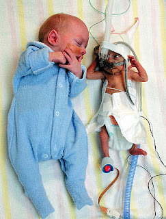
Twins delivery, with one twin 3 times larger than the other one


Byron and Lincoln Ryman was born in March 2007. Their parents, Todd and Nicole Ryman, have been trying to have kids for so long, and finally Mrs Ryman got pregnant through in vitro fertilization (IVF), and she got a twin pregnancy.

Later on, the doctor noticed that there were some problems with her pregnancy. It was discovered that one of the twins was significantly smaller than the other one. The twins developed twin to twin transfusion syndrome, which may occurs only to some fetuses who shared the same placenta (monochorionic twins). The unbalanced transfusion between the babies cause Bryon to get hyper perfused while Lincoln was getting hypo perfused.

The parents were given an option to terminate Lincoln, the smaller baby, in hopes of saving Bryon. However, they didn’t agree. “I wanted to have 2 babies not one”, said Mrs Ryman.

During the pregnancy she was having weekly scans and she said the doctors would ‘write Lincoln off’ and told her to ‘be prepared to not see a heartbeat next week’.

In March 2007, she was induced at 29 weeks of gestation after the doctor discovered that Bryon had stop growing, and delivery of the babies seems to be the best option. The babies were born 11 weeks premature. Bryon, the firstborn is 3 times larger than his little brother, Lincoln. Bryon birth weight was 1.54kg while Lincoln was 530g only.

Born prematurely, both babies had been struggles to survive those critical periods with all the complication of premature birth. Initially the parents thought that they will have two boys with multiple health issues. In the beginning they had suffered from breathing problems, heart condition, and Lincoln had to take his food through feeding tube until the age of two. However, as miraculous as they are, these boys condition improved progressively and are doing very well.


________________________________________________________

http://www.godvine.com/They-Were-Told-to-Abort-One-of-their-Twins-But-Watch-This-1523.html
CLICK TO WATCH

________________________________________________________